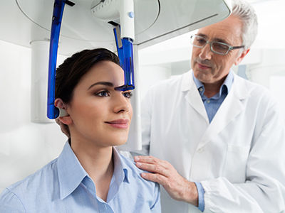
At the office of Today's Dentistry San Jose, we combine thoughtful clinical judgment with advanced imaging tools to make diagnosis and treatment more predictable. One of the technologies that has reshaped modern dentistry is cone-beam computed tomography (CBCT). By producing three-dimensional images of the teeth, jaws, and surrounding structures, CBCT gives our clinicians a richer view of anatomy than conventional two-dimensional X-rays can provide.
Our practice invests in a high-resolution CBCT system that balances image clarity with patient safety. The result is detailed diagnostic information in a quick, comfortable scan that supports more accurate treatment planning across restorative, surgical, and orthodontic care. Below, we explain what CBCT is, how it’s used, and why it matters for patients seeking precise, modern dental care.
Traditional dental X-rays are excellent for many routine evaluations, but they are inherently flat. CBCT captures volumetric data, which means we can assess depth, spatial relationships, and fine anatomical detail in three planes. For clinicians, that extra dimension reduces uncertainty: what might look ambiguous on a two-dimensional film often becomes clear on a CBCT scan.
This improved visualization helps detect conditions that could otherwise be missed or misjudged, such as small fractures, complex root anatomy, localized infections, or bone defects. Because the images are distortion-free and true-to-scale, they give us a reliable foundation for diagnoses that guide subsequent treatment decisions.
For patients, the practical benefit is consistent care planning. When we can see the full picture of a problem, we can explain it more clearly, propose options with greater confidence, and reduce surprises during treatment. In short, CBCT supports smarter clinical choices and clearer communication between patient and provider.
CBCT produces a three-dimensional dataset that captures hard tissues in exceptional detail. This means clinicians can examine tooth positions, the thickness and height of the jawbone, the course of nerve canals, and the relationship between the sinuses and upper teeth — all from a single, comprehensive scan. Those insights are especially important in areas where anatomy is complex or varies significantly from patient to patient.
Beyond routine structure, CBCT can reveal subtle anatomical variations that affect treatment risk and outcome. For example, the exact pathway of the inferior alveolar nerve or the presence of a hidden root canal branch can change how a procedure is approached. By identifying these factors in advance, the care team can adapt techniques and reduce the likelihood of complications.
CBCT also helps in screening for certain bony pathologies and developmental anomalies. While not a replacement for a clinical examination or other diagnostic tests, the scan is a powerful supplement that clarifies the underlying anatomy and highlights issues warranting further evaluation or monitoring.
One of the most transformative applications of CBCT in dentistry is in implant planning. Successful implant placement depends on precise knowledge of bone volume, density, and nearby anatomical structures. A three-dimensional scan lets us map implant sites virtually, choose properly sized implants, and determine ideal angulation before entering the operatory.
CBCT-based planning reduces guesswork and allows for the fabrication of surgical guides when indicated. These guides help translate the virtual plan into exact clinical placement, which can shorten surgical time and improve restorative outcomes. For patients, that means a more predictable process and better long-term function for implant-supported teeth.
Beyond implants, CBCT supports complex extractions, evaluation of impacted teeth, and the approach to jaw surgery. By informing preoperative decisions with detailed imagery, clinicians can minimize intraoperative surprises and tailor surgical strategies to each patient’s unique anatomy.
Safety is a central consideration when choosing imaging modalities. Modern CBCT units are designed to capture high-quality images with focused fields of view and optimized exposure settings that limit radiation to the area of interest. When used selectively and with appropriate protocols, CBCT delivers diagnostic value while maintaining a conservative radiation profile.
Scans are typically fast — often completed in less than a minute — and the patient experience is straightforward and noninvasive. The machine’s open design and brief scan time make it more comfortable for many patients than other imaging options that require constrained positioning or longer exposures. Our team follows ALARA principles (as low as reasonably achievable) to ensure imaging is justified and tailored to each clinical question.
Before recommending a scan, we review the expected benefits relative to any potential risks and discuss the rationale with patients. This transparent approach helps patients understand why a CBCT scan may be the most appropriate diagnostic step for their care.
A CBCT scan is only as useful as the clinical interpretation that follows. Our clinicians combine radiographic findings with clinical examination, patient history, and treatment goals to form a comprehensive plan. We review the scan in detail, identify relevant findings, and explain how those observations influence recommended therapies.
When necessary, we collaborate with specialists — oral surgeons, endodontists, periodontists, and radiologists — to ensure complex findings are evaluated from multiple perspectives. The three-dimensional data set can be exported and shared securely with consulting clinicians to support coordinated care and consistent treatment planning.
CBCT also integrates with digital workflows used for restorative design and surgical guide fabrication. When a virtual plan is translated into a physical guide or a laboratory restoration, the accuracy of the scan helps deliver predictable, well-fitting results. Ultimately, the technology strengthens the link between diagnosis and execution, improving both function and long-term success.
In summary, cone-beam computed tomography is a powerful diagnostic tool that enhances precision across many areas of dental care. At Today's Dentistry San Jose, we use CBCT selectively and thoughtfully to support clearer diagnoses, safer surgeries, and more predictable restorative outcomes. If you would like to learn more about how CBCT might apply to your treatment, please contact us for additional information.

CBCT is a three-dimensional imaging technology that captures volumetric X-ray data of the teeth, jaws, and surrounding structures in a single scan. The machine rotates around the head to create a detailed dataset that practitioners can view in axial, coronal and sagittal planes. Because the images are true to scale, clinicians use them to visualize depth and spatial relationships that are not apparent on standard two-dimensional X-rays.
The resulting dataset is typically reconstructed as cross-sectional slices and 3D renderings that reveal bone contours, tooth roots, nerve canals and sinus anatomy. These images support more precise diagnoses and treatment planning across restorative, surgical and orthodontic care. CBCT is a supplement to, not a replacement for, a clinical exam and conventional radiography when those methods suffice.
Traditional dental X-rays produce flat, two-dimensional images that are excellent for routine evaluations of cavities and basic tooth structure. By contrast, CBCT acquires a volumetric dataset that provides depth information and distortion-free measurements, which are essential when anatomy is complex or when three-dimensional insight affects treatment decisions. The difference is analogous to comparing a photograph with a small volumetric scan of the same area.
CBCT units also allow clinicians to select different fields of view and resolution settings to focus on a localized area or capture a broader region. For many routine tasks, periapical or panoramic X-rays remain appropriate because they involve lower exposure and faster processing. Clinicians reserve CBCT for cases where the additional anatomical detail will meaningfully influence diagnosis or surgical planning.
CBCT is commonly indicated for implant planning, evaluation of impacted or atypically positioned teeth, assessment of complex root anatomy for endodontic cases, and analysis of jaw pathology or trauma. It is also helpful for evaluating temporomandibular joint relationships and for orthodontic cases that demand detailed skeletal assessment. In each situation, three-dimensional images reduce uncertainty and help determine the safest and most effective approach.
Clinicians may order a CBCT when standard radiographs leave questions about bone volume, nerve location, or hidden anatomical variations that could change the risk profile of a procedure. The decision to scan is based on the clinical question being asked and whether the additional information will alter treatment. When CBCT is not expected to change management, less complex imaging is typically chosen.
For implant planning, CBCT provides accurate information about bone height, width and density as well as the proximity of the inferior alveolar nerve and maxillary sinuses. This allows clinicians to select implant length and diameter and to determine ideal angulation without relying on estimation. Virtual implant placement within the scan can reveal whether bone augmentation or alternative approaches are needed before a surgical procedure begins.
CBCT data can also be used to fabricate surgical guides that translate virtual plans into precise clinical placement, which often reduces operative time and improves prosthetic outcomes. For extractions, evaluation of impacted teeth and corrective jaw procedures, the ability to anticipate anatomic challenges decreases intraoperative surprises. Overall, three-dimensional planning contributes to safety, efficiency and predictable restorative results.
Modern CBCT units are engineered to minimize radiation by using focused fields of view and adjustable exposure parameters, and clinicians adhere to ALARA (as low as reasonably achievable) principles when recommending scans. In many dental applications, CBCT involves lower radiation than conventional medical CT while delivering the specific anatomic detail needed for dental planning. The technology is used selectively and only when the diagnostic benefit outweighs any potential risk.
Before ordering a scan, the care team will explain why CBCT is being recommended and how the information will influence treatment. Patients with specific concerns about radiation or special health circumstances should discuss them with the clinician so the team can tailor imaging choices and protective measures appropriately. Pregnant patients and those with recent equivalent imaging should always inform the office so clinicians can consider alternatives when appropriate.
A typical dental CBCT scan is quick and noninvasive, often completed in under a minute once the patient is positioned. Patients may sit or stand while the scanner rotates around the head, and the process usually requires only that metal objects such as eyeglasses or removable dental appliances be removed. The open design and short scan time make the experience more comfortable than many other imaging procedures.
There is no injection or contrast required for routine dental CBCT, and most people are able to remain still for the brief duration of the acquisition. After the scan, the images are reconstructed and reviewed by the clinician, who will discuss the findings and explain how they affect the proposed treatment plan. If additional specialist review is needed, the dataset can be shared securely with consulting clinicians.
Interpreting CBCT requires correlating radiographic findings with the clinical exam, patient history and treatment goals to form a comprehensive plan. Clinicians evaluate cross-sectional slices and three-dimensional renderings to identify relevant anatomic features, pathology, or risk factors that influence procedural choices. When findings are complex, collaboration with specialists such as oral surgeons, endodontists or oral radiologists helps ensure a well-rounded assessment.
CBCT data are also compatible with digital workflows used for restorative design and surgical guide fabrication, enabling accurate communication between the clinic and dental laboratories or guide manufacturers. Exportable DICOM files allow for virtual planning and verification before any irreversible steps are taken in the operatory. This integration improves predictability and helps align surgical execution with the restorative objective.
While CBCT excels at visualizing hard tissues, it provides limited contrast for soft tissues and is susceptible to artifacts from metal restorations and patient movement. These limitations can reduce diagnostic clarity in certain scenarios and may necessitate complementary imaging or referral for alternative studies when soft-tissue detail or artifact-free imaging is essential. CBCT is not a universal replacement for all radiographic exams and should be chosen for specific clinical questions it can answer well.
Other practical limitations include field-of-view constraints and variations in image quality between different units, so clinicians select scan parameters carefully to balance diagnostic needs and exposure. Additionally, not all findings on a CBCT scan represent active disease; some observations require clinical correlation or follow-up imaging. Clear communication about what the scan can and cannot show helps manage expectations and guide next steps.
CBCT datasets provide the three-dimensional foundation for virtual planning tools that align implant position with the final prosthetic design, enabling prosthetically driven implant placement. When combined with intraoral scans or digital impressions, CBCT data allow technicians and clinicians to design restorations and surgical guides that fit accurately within a patient’s anatomy. This digital coordination reduces manual guesswork and streamlines the transition from planning to clinical execution.
Surgical guides produced from CBCT-based plans help translate virtual placements into reproducible clinical results by controlling drill trajectory and depth during implant osteotomy. The result is often improved precision, predictable restorative emergence profiles and more efficient surgical appointments. Integration with CAD/CAM workflows also facilitates communication between the dental team and external laboratories or milling centers.
Patients should ask about CBCT when undergoing implant consultations, when symptoms or prior imaging suggest complex root anatomy or pathology, or when a clinician indicates that three-dimensional detail will change treatment decisions. It is also appropriate to discuss CBCT before surgical procedures involving impacted teeth, bone grafting or corrective jaw work. Open discussion helps ensure that imaging is used judiciously and tailored to the clinical question at hand.
At Today's Dentistry San Jose, our team reviews each case individually and explains why a CBCT scan is recommended or why alternative imaging may be sufficient. We discuss the diagnostic benefit, the imaging process and any necessary precautions so patients understand how the scan supports their care. If you have specific concerns or questions about imaging, please raise them during your consultation so the team can address them directly.

Ready to schedule your next dental appointment or have questions about our services?
We’re ready whenever you are. Whether you have a quick question about a treatment or you're set to book your next visit, our team makes getting in touch effortless. Give us a call, drop an email, or use the form below to start a conversation. Your path to a healthier, more confident smile is just a message away—reach out today and let’s discuss how we can help.