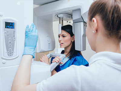
The image shows a person receiving medical treatment with a device attached to their face, while another individual is using a microscope to observe them closely.

X-Ray Dentistry in San Jose

What digital radiography is and why it matters

Digital radiography replaces traditional film with electronic sensors and computer processing to capture dental X-ray images. Instead of exposing film and developing it in chemicals, a digital sensor or plate records the image data and sends it immediately to a computer for review. This evolution in imaging has changed how clinicians diagnose, plan, and communicate about oral health by delivering higher-quality images faster and with fewer steps.

For patients, the practical benefits are easy to see: appointments can move more efficiently, images are available for discussion during the same visit, and clinicians can manipulate contrast and zoom without having to retake films. For the dental team, digital files are simpler to archive, retrieve, and share securely with specialists when collaborative care is necessary. That combination of efficiency and clarity is why digital radiography is now a standard of care in modern dental practices.

Because it integrates seamlessly with electronic health records, digital radiography also supports continuity of care. Images become part of a permanent, searchable record that helps monitor changes over time, supports long-term treatment planning, and reduces the chance of lost or damaged film. In short, digital radiography improves diagnostic capability while simplifying recordkeeping.

How sensors and plates capture sharper images

Digital sensors come in several forms, most commonly wired or wireless intraoral sensors and phosphor storage plates (PSPs). Each device captures X-ray photons and converts them into an electronic signal that a computer translates into the visual image you see on the screen. The result is a crisp, high-resolution image that can be magnified and adjusted to reveal subtle anatomical details that film might miss.

Because the image is created digitally, clinicians can apply software tools to enhance visibility—adjusting brightness, contrast, and edge detail to better evaluate cracks, early decay, or the margins of restorations. These adjustments are nondestructive, meaning they don’t alter the original file and can be reversed or saved as separate views for comparison over time. This flexibility improves diagnostic confidence without additional radiation exposure.

In practice, this means fewer retakes and a clearer diagnostic pathway. When clinicians can see fine differences in density and structure, they are better equipped to detect early-stage problems and recommend timely, conservative care. The enhanced image quality of digital systems supports both routine exams and more complex evaluations, such as assessing root anatomy prior to endodontic treatment.

Radiation safety: understanding dose and protection

One of the most important advantages of digital radiography is its ability to produce diagnostic images at a lower radiation dose than traditional film. Advances in detector sensitivity and image-processing algorithms allow useful clinical images to be generated with less exposure. While any X-ray involves some level of radiation, modern dental imaging is tightly controlled and designed to keep doses as low as reasonably achievable.

Dental teams follow established safety protocols to protect patients, including the use of lead aprons when appropriate, collimation to limit the beam to the area of interest, and strict adherence to exposure settings tailored to each patient. These measures, combined with digital detectors, reduce unnecessary exposure while still delivering the information clinicians need to make informed decisions about care.

Patient concerns about radiation are understandable, and transparent communication helps address them. Clinicians can compare digital dental doses to familiar background radiation sources to provide context and will only recommend imaging when it directly contributes to diagnosis or treatment planning. In routine care, digital radiographs offer a favorable balance of diagnostic benefit and patient safety.

From capture to care: how images support treatment planning

Digital images become powerful tools when integrated into the treatment workflow. After capture, images are immediately available for review alongside medical and dental histories, intraoral photographs, and other diagnostic data. This convergence of information helps clinicians craft personalized treatment plans and explain options clearly during the appointment, improving understanding and shared decision-making.

Because images can be enlarged and annotated in real time, clinicians can point out areas of concern and walk patients through what they see — whether that’s the start of a cavity, bone loss near a tooth, or the fit of a crown preparation. When specialists are involved, digital files can be shared securely, accelerating referrals and ensuring that outside providers have precise visual data to guide their interventions.

Digital image archives also support follow-up care. Comparing current and prior images makes it easier to monitor healing after procedures, assess the progression of conditions, and evaluate the long-term success of restorations or implants. This longitudinal perspective is a key advantage of keeping structured, accessible digital records.

Environmental and operational benefits beyond the operatory

Switching to digital imaging reduces the environmental footprint associated with radiographic processing. Because no chemical developers or fixer solutions are required, practices avoid hazardous waste and the additional handling and disposal that film processing demands. Fewer consumables and less physical storage also lower ongoing operational burden.

Operationally, the ability to retrieve an image instantly improves throughput and patient experience. Multiple care team members can view the same image at once on separate monitors, facilitating collaboration during complex visits. Digital images also simplify insurance documentation and case presentation, since files can be exported, printed, or embedded in treatment notes without re-photographing or scanning physical films.

For practices committed to continuous improvement, digital radiography is more than a single piece of equipment — it is a platform that supports other technologies, such as digital impressions and three-dimensional imaging. Together these systems create a cohesive digital ecosystem that elevates diagnostics, treatment planning, and patient communication.

In summary, digital radiography is a major advance in dental imaging that combines clearer images, faster workflows, and enhanced patient safety. The technology supports better diagnostics, simplifies recordkeeping, and reduces environmental impact, all while integrating into modern digital dental ecosystems. If you have questions about how digital radiography is used in our practice or how it might affect your care, please contact us for more information.

The image shows a logo with text that reads  TD  at the top, followed by  Today s Seniors Testimony,  indicating that the logo likely represents an organization related to senior citizens sharing their experiences.

Frequently Asked Questions

What is digital radiography and why does it matter?

+

Digital radiography replaces traditional film with electronic sensors and computer processing to capture dental X-ray images. A sensor or phosphor plate records X-ray information and sends it to a computer for immediate review. This digital workflow eliminates chemical development and delivers higher-resolution images quickly.

Those faster, clearer images help clinicians diagnose conditions earlier and explain findings to patients during the same visit. Digital files are easy to archive, retrieve, and share with specialists when collaborative care is needed. At Today's Dentistry San Jose, we use digital radiography to streamline visits and support precise treatment planning.

How do digital sensors and plates capture sharper images?

+

Digital sensors typically include wired or wireless intraoral detectors and phosphor storage plates (PSPs) that capture X-ray photons and convert them into electronic signals. The detector’s electronics and software translate those signals into a high-resolution image that can be displayed and manipulated on a monitor. This conversion produces sharper detail than traditional film and enables magnification without loss of information.

Because images are recorded digitally, clinicians can apply nondestructive enhancements such as brightness, contrast, and edge sharpening to reveal subtle anatomy. Adjustments can be saved as additional views for comparison while preserving the original file. That flexibility reduces the need for retakes and improves diagnostic confidence.

Is digital radiography safe in terms of radiation exposure?

+

Digital radiography generally requires less radiation than traditional film because modern detectors are more sensitive and software can produce usable images from lower exposures. Dental teams adhere to the principle of keeping doses as low as reasonably achievable and tailor exposure settings to each patient's size and diagnostic need. Protective measures such as collimation and lead aprons are used when appropriate to further limit exposure.

Patient questions about radiation are common, and clinicians can explain relative doses and why an image is recommended for diagnosis or treatment planning. Imaging is only advised when the expected diagnostic benefit outweighs the small amount of exposure. For routine exams and follow-up care, digital radiographs provide a favorable balance of diagnostic value and safety.

How does digital radiography improve diagnosis and treatment planning?

+

Immediate availability of digital images transforms how clinicians plan care and communicate with patients. Images can be annotated, measured, and shown next to medical history and intraoral photographs so patients understand findings and treatment options. This visual approach supports shared decision-making and helps clinicians recommend conservative, timely interventions.

Digital images also support long-term monitoring by making it easy to compare current and prior views for subtle changes over time. When specialists are involved, files can be shared securely to accelerate referrals and ensure everyone has precise visual data. That continuity reduces diagnostic delays and supports coordinated care.

Will my digital images become part of my dental record and how are they stored?

+

Digital radiographs become part of the patient's clinical record and are stored electronically alongside treatment notes and other diagnostic files. Electronic storage simplifies retrieval for future visits and reduces the chance of lost or damaged films. Records can be indexed and searched, making longitudinal care and trend analysis more practical.

Dental practices maintain secure systems and access controls to protect patient images and associated data. When images are shared with outside providers, secure transmission protocols are used to preserve privacy and integrity. Patients can request copies of their images or have them sent to specialists when necessary for coordinated care.

Can digital radiography reduce the need for retakes and additional imaging?

+

High-resolution digital images and real-time software adjustments reduce the frequency of retakes by allowing clinicians to correct exposure and contrast on the spot. Improved detector sensitivity and immediate feedback help the team capture diagnostically useful images the first time. This efficiency shortens appointments and minimizes additional radiation.

Despite these advantages, two-dimensional digital radiographs have limits for certain clinical questions, such as complex root anatomy or detailed bone structure. In cases that demand three-dimensional detail, clinicians may recommend supplemental imaging like cone beam CT for a fuller view. Those decisions are made based on the specific diagnostic need and the potential benefit to the patient's treatment plan.

How does digital radiography support specialized treatments such as implants and root canals?

+

Digital radiography plays an important role in planning treatments such as dental implants and endodontic therapy by revealing tooth and bone anatomy. Precise two-dimensional images help assess root morphology, periapical health, and the relationship of teeth to adjacent structures. When combined with other digital tools, these images support predictable, conservative decision-making.

For implant placement and complex surgical planning, clinicians often integrate two-dimensional radiographs with three-dimensional scans and digital impressions to create a comprehensive treatment plan. This layered approach enables accurate measurements and prosthetic planning while minimizing surprises during surgery. The interoperability of digital systems improves coordination between restorative and surgical teams.

What environmental and operational benefits come from switching to digital imaging?

+

Switching to digital imaging eliminates the need for chemical developers and fixers, reducing hazardous waste and the environmental impact of film processing. Digital workflows also decrease the use of consumables and the physical storage space required for films. These operational benefits align with broader sustainability goals while simplifying daily practice management.

Operationally, instant image access improves patient throughput and allows multiple team members to review the same image simultaneously. Digital files streamline insurance documentation and case presentation by making exports and annotations straightforward. Over time, the reduced material burden and faster workflows can improve practice efficiency and patient experience.

What should I expect during a digital X-ray appointment?

+

During a digital X-ray appointment, a small sensor or plate is positioned in the mouth and the patient is asked to hold still while a brief exposure is taken. The capture is quick, often completed in seconds, and most patients experience minimal or no discomfort beyond mild pressure from the sensor. Images appear on the computer immediately, allowing the clinician to confirm quality and repeat a shot only if clinically necessary.

Clinicians will explain what the images show and how they factor into diagnosis or treatment planning, answering any patient questions in real time. Special accommodations are available for patients with strong gag reflexes, limited jaw opening, or mobility issues to make the process more comfortable. If additional imaging is needed for complex cases, the clinician will explain the reason and the next steps.

How are my images protected and how can I share them with other providers?

+

The practice stores and manages patient images using secure electronic systems with controlled access to protect privacy and maintain integrity. Encryption, password protection, and regular backups are common safeguards that limit unauthorized access and preserve files for clinical use. Staff training and administrative procedures further support proper handling of diagnostic images.

If patients have questions about how their images are used or wish to share them with another provider, the dental team can explain the available options and obtain appropriate authorizations. Electronic transfers are conducted using secure methods to preserve confidentiality and ensure specialists receive the necessary diagnostic information. For more specific inquiries about imaging policies or record access, patients may contact Today's Dentistry San Jose directly.

A woman sitting on a dental chair receiving treatment from a dentist while another person observes.

Get in Touch with Today's Dentistry San Jose For Exceptional Dental Care

Ready to schedule your next dental appointment or have questions about our services?

We’re ready whenever you are. Whether you have a quick question about a treatment or you're set to book your next visit, our team makes getting in touch effortless. Give us a call, drop an email, or use the form below to start a conversation. Your path to a healthier, more confident smile is just a message away—reach out today and let’s discuss how we can help.